Research Themes
Context and challenges
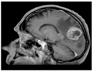
With a rising incidence up to 5 new cases each year per 100,000 inhabitants (13000 new cases per year in Europe), glioblastoma (GBM) is the third cause of death by cancer in young adults population (aged from 15 to 34). It is an incurable tumour whose median survival is less than 18 months. This oncology domain remains of great interest in both preclinical and clinical research. The standard treatment is based on surgical resection followed by brain irradiation associated with chemotherapy.
The goal of surgery is to achieve maximum reduction in tumour volume without affecting the functional outcome of patients according to their pejorative prognosis. Nevertheless, even when surgical resection is complete, the invasiveness of glioblastoma and its proliferating nature do not allow a local control with standard therapeutic protocols. Indeed, patients whose tumour location is not eligible for surgery have limited survival.
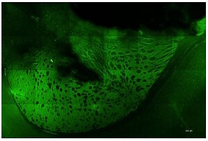
Photodynamic therapy (PDT) consists in exposing photosensitized tumour cells to laser light. Local or general administration of a pharmacological agent (precursor) is previously required to allow photosensitization of the cells. Our research focus on the 5-Aminolevulinic Acid (5-ALA), which induces a relatively specific photosensitizer accumulation: protoporphyrin IX (PPIX). The photosensitizer is present only in tumour cells and leads to their specific destruction upon illumination, while sparing healthy tissues.
The NeuroPDT research program developed by ONCOTHAI aims at developing an effective, reproducible and selective pattern for photodynamic therapy using 5-ALA. Nowadays, the on-going preclinical studies are confirming the efficiency of the 5-ALA laser therapy while revealing the harmlessness of its delivery. As a clinical approved molecule of 5-ALA for fluorescence guided resection, this therapy could be rapidly transferred to the clinical practice.
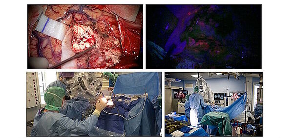
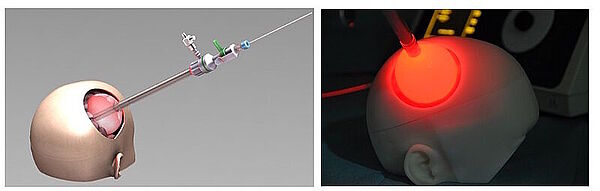
Using photodynamic therapy, the light exposure of photosensitized cells can be achieved according to two different patterns: exposure to red light of the operative site during surgery or insertion in the brain of optical fibers, for treatment distant from surgery. The latter solution could also represent an alternative therapeutic option for patients with inoperable or recurrent glioblastoma.
Eventually, the expected results would lead to a significant improvement in patients’ overall survival subsequently to a selective tumor reduction, assessed by MRI.
Goals
The purpose of the project is to propose two distinct treatments patterns: intraoperative photodynamic therapy (intraoperative PDT) and interstitial photodynamic therapy (iPDT). Indeed, these two modalities are complementary since intraoperative PDT is a promising adjuvant treatment to enhance the current therapeutic armamentarium (surgery, radiotherapy and chemotherapy) and iPDT is a new modality for the treatment of non-operable glioblastomas.
Integrating these new treatments in the management of the patient requires several research stages: preclinical study - clinical studies - methodological developments.
A pre-clinical study was conducted in the laboratory to validate the assumptions of efficacy and optimize the PDT treatment strategy.
Optical fibres coupled to a laser diode allowed a focused illumination of the tumour site in vivo. Thus, we were able to assess the response of glioblastoma treatment PDT. On another level, this study allowed us to optimize treatment modalities, controlled all the workflow steps (planning - treatment - monitoring) and finally validated the proof of concept of PDT.
The methodological findings concern both the image processing and treatment scheme for iPDT or intraoperative PDT.
Today, a comprehensive study of imaging techniques is implemented to identify the best solutions for planning, guiding and monitoring the treatment. The imaging panel covers both the metabolic imaging by Positron Emission Tomography (PET) and MRI.
In this context, PET images segmentation algorithms are developed to accurately quantify the metabolic activity of the tumor. This step is crucial because PET imaging combined with MRI images will ensure the definition of tumor invasion limits and help to adapt the treatment pattern, to get a better target for the therapy.
Where do we stand?
Clinical studies aim to validate all the implemented developments to deliver the optimal treatment.
Today, a single-center clinical trial is underway at the Lille University Hospital to demonstrate the feasibility and the safety of intraoperative PDT. This study, INDYGO (Intraoperative photodynamic therapy of glioblastoma) aims to deliver photodynamic therapy during surgery glioblastoma with a device developed in the laboratory.
On the international side, ONCOTHAI coordinates a European Network: SYNAPS (Synergizing Photodynamic Therapies for Neurosurgery). The network relies on a partnership between neurosurgical departments, research centres and health technologies companies. The objective of SYNAPS is to achieve a multicenter clinical trial and randomized across Europe.
Finally, this project will remove the constraints of using this technique for the treatment of these still incurable tumours. PDT laser treatment could quickly complete the therapeutic armamentarium for glioblastoma, and even become a new therapeutic option for inoperable tumour locations.
Topic
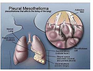
Malignant pleural mesothelioma (MPM) is an aggressive serosal tumor of the pleura. Its main aetiological agent is an exposure to asbestos fibers, mostly work-related, and the disease appears after a latency of 30 to 40 years after initial exposure. MPM is considered as a rare tumor: in France, its incidence is 900 cases every year. However, its incidence is rising throughout the world because of the increasing use of asbestos until the 1970’s and will peak in the next decade. We also fear a pandemic rise of MPM in the future with developing countries (China, Brazil, India…) still using asbestos today. MPM has a poor prognosis with a median survival of less than a year. This can be explained by the delay of diagnosis due to late symptoms, with a disease already advanced locally, the difficulty of obtaining a confident anatomopathological diagnosis, and a complex treatment with deceiving outcomes.
Treating MPM remains a challenge. MPM shows a strong resistance to chemotherapy, which is often the unique treatment available for patients with advanced and unresectable disease, or who cannot sustain a heavy thoracic surgery, with a median survival of 12,1 months. Surgery offers the best chance of survival for this still incurable disease. However, after the most complete tumor resection, microscopic tumor cells persist and surgery should be associated with a local adjuvant treatment. Up to this day, there are no international recommendations on surgical treatments with a curative intent. Surgery is still investigational and should be part of a multimodality treatment and included in a prospective clinical trial. In January 2012, a clinical French network of regional expert centres for the care of MPM was created: “MESOCLIN”. It is financed by the National Institute against Cancer or INCa. This network is coordinated par the expert centre of Lille, in close cooperation with “Mesopath”, a French anatomopathological expert network.
Medical interest for this cancer has evolved during these last years, with a real investment in its treatment: with the notion of reasonable surgery, with relatively efficient cyto-toxic drug combination, with the emergence of biotherapy and new radiotherapy modalities, and especially the development of concerted multidisciplinary strategies which allow to build randomised, prospective clinical trials, that had been lacking on this field. The recruitment of patients with MPM is important in Lille, due to strong professional and environmental exposure to asbestos fibres in the “the Hauts de France” area: shipyards, metallurgy, building industry…
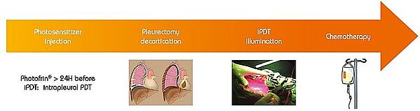
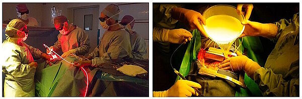
Photodynamic therapy is a treatment of ancient principle, but is relatively new in the multimodal management of MPM. The effect of PDT requires the interaction of 3 components: a photosensitizer (PS), oxygen in the tissue and light with the specific wavelength activating the PS. None of them are individually toxic but when put together they induce a tumoricidal photochemical reaction. When tumour cells absorb the light, the photosensitizer produces free radicals and oxidation reactions which lead to the death of the treated cells and kill tumour angiogenesis. Intrapleural PDT is based on a two-stage process: intravenous administration of a photosensitizer with a specified drug dose and drug-light interval before surgery, and intraoperative illumination of the pleural cavity, after maximal resection of the tumor, by a laser source at an appropriate wavelength, and at a specified light dose. The appeal of PDT as an adjuvant treatment relies on its relative tumor selectivity, depending on a photosensitizer able to direct itself and remain longer in the tumor cells than in the normal cells, and an illumination restricted to the cancer superficial zone.
For a few decades, photodynamic therapy has been the subject of many studies (especially phase I and II studies) as part of a multimodal treatment of malignant pleural mesothelioma, including extra pleural pneumonectomy (EPP) or pleurectomy decortication (P/D). If preliminary results were disappointing, two recent studies by Friedberg (USA) have demonstrated that Photofrin®-mediated PDT, after complete macroscopic resection, and associated with cisplatin based chemotherapy, offers a remarkable survival with reasonable toxicity. Even in patients with locally advanced stages of MPM, and when a lung-sparing procedure (P/D) is performed, survival was much higher than without adjuvant PDT: the median OS (Overall Survival) was 31,7 months (95% [CI] 9-54,3 months).
Goals
ONCOTHAI team, in collaboration with the departments of Pulmonology and Thoracic Oncology, and Thoracic Surgery, in Lille University Hospital, have started an ambitious project aiming at developing intrapleural PDT for MPM.
On the clinical side, the objective was to combine intrapleural PDT to P/D and adjuvant chemotherapy, for patients with MPM. There was already a close connection between the two teams of France and USA, who met many times. The idea was to reproduce identically the surgical procedure and the PDT performed by the team of Philadelphia, thanks to their collaboration.
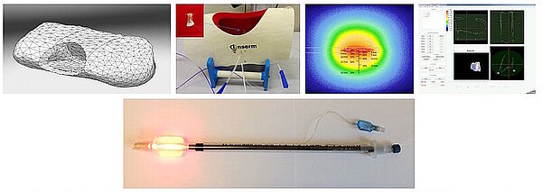
On the research side, the objective was to optimize the illumination through the light device and the development of an alternative intra operative dosimetry method for intrapleural PDT. Complete and uniform illumination of the pleural cavity is very difficult to obtain, due to its complex geometry and the presence of surrounding « noble » organs. The actual technique consists in filling the cavity with diffusing solution and moving the light device around the chest cavity. Today, light dosimetry is achieved using seven sensors collecting light dose and fixed inside the pleural cavity at strategic locations, but it does not give information about the light delivered between these points. The research progamme of INSERM U1189 is based on skills, already used for other pathologies: laser action modelling and medical imaging. The aim is to optimize light dosimetry and ensure complete and uniform light delivery to the targets, which is the key to the success of PDT.
Where do we stand?
With the support of the Regional Board of Nord Pas de Calais ans patients associations, a local phase II clinical trial is today ongoing at the University Hospital of Lille. Then, a multicentric randomized phase II trial will take place in France, to validate the benefit of PDT for MPM. This second clinical trial takes place within a Clinical Research Hospital Programme (PHRC) selected by the National Cancer Institute (INCa).
On the experimental side, an illumination profile of a light device has been defined and combined with an electromagnetic spatial tracking system, with 3D imaging (chest CT scan) visualization of of the light dose delivered in real time. This new dosimetry approach has been validated on a intraoperative hemithorax phantom, conceived and 3D printed within the laboratory.
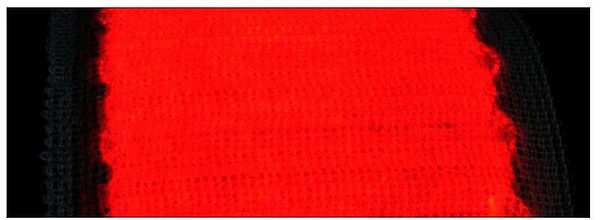
Another path of research on which INSERM U1189 is working is the use of flexible light textile which could well be applied on the pleural cavity walls. Recent work, has demonstrated that flexible light textile could ensure homogenous light delivery on a peritoneal carcinomatosis animal model.
Topic
Actinic keratoses (AKs) are erythematous and squamous skin lesions, rough to the touch, mainly present on sun-exposed areas. AKs are common; their prevalence in the U.S. and Europe is estimated between 11 and 25% of the population. In France, they concern 754/100.000 patients and represent the first cause of consultation in dermatology.
They are more likely to reach the elderly, with a clear phototype, who worked outside. Sometimes painful and hemorrhagic on contact, these lesions may evolve towards an invasive squamous cell carcinoma. AKs are often multiple. The apparently healthy skin between lesions presents abnormalities in histology and in biological analysis (leading to the notion of “field cancerization”).
It is estimated that for an individual with 7.7 AK, the probability that at least one of these lesions progresses to invasive squamous cell carcinoma over a period of 10 years is 10%. Malignant potential and the unpredictable evolution of AKs led to a consensus on the need to treat.
Photodynamic therapy (PDT) relies on the exposure to a light of a particular wavelength of photosensitized tumor cells. In dermatology, the photosensitization is given by the topical application of methyl aminolevulinate (MAL) contained in the cream Metvixia® (Galderma SA). Applied on the area to be treated, this pharmacological agent induces a specific accumulation of the photosensitizer Protoporphyrin IX (PpIX) in the tumor cells.
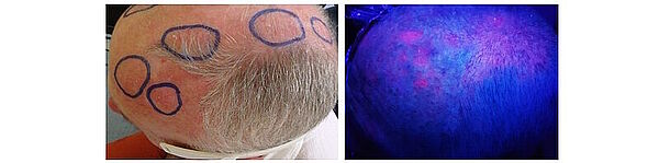
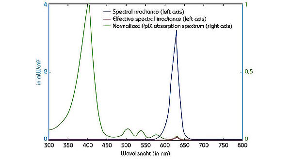
The light source most commonly used in Europe to activate the PpIX is the Aktilite CL 128 lamp (Galderma SA). This lamp, which is a rigid LEDs panel, emits a red light at a wavelength of 630 nm and delivers a light dose of 37J/cm² (75 mW / cm²).
Although often described as very painful by patients, PDT remains a first line treatment for the management of multiple AKs because it allows to treat large areas with a satisfactory response rate and excellent cosmetic results.
Goals
DermatoPDT program aims to develop new illumination devices and protocols, which should partially solve these limitations.
Gathering technical and software platforms, the program includes in particular research on the mathematical modeling1 of the interaction between light and biological tissue, and the development of softwares allowing the analysis and comparison of light sources2.
The DermatoPDT research program is also based on the development of new devices, integrating new illumination modalities3.
Within the framework of project ANR-12-EMMA-0018, the ONCOTHAI unit contributed to the development of a new illumination device: Flexitheralight4.
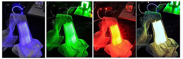
Based on the use of a light emitting fabric, conformable to the complexity of human anatomy, it guarantees a homogeneous illumination on all the lesions (especially on the scalp). In addition, light emitting fabric diffuses a red light (635 nm), with a low fluence rate (12 mW / cm²) in order to minimize the pain usually reported by patients.
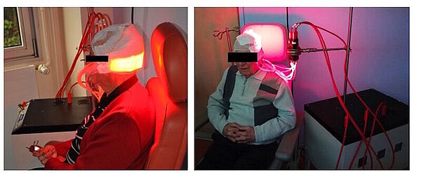
In 2013, the ONCOTHAI team initiated the European project CIP 621103 Phosistos, and its partnership with various laboratories and manufacturers throughout Europe, resulted in the development of a new device integrating a mature light emitting fabric technology, more functional and industrializable5.

Where do we stand?
In 2016, Phosistos6 was evaluated in the management of AKs in a comparative, intra-individual, randomized phase II study conducted in the dermatology departments of Lille University Hospital in France, and Klinikum Vest in Germany. The main hypothesis of this study was the safety of both Phosistos device and illumination modality, the non-inferiority in terms of efficiency and the better tolerance compared to conventional PDT.
About forty patients with at least 10 AKs were included in this study. The included lesions were divided into 2 groups: the first group received the conventional PDT (with the rigid panels and a high fluence rate: 75 mW/cm²) while the second group was treated with the Phosistos device. The 3-month therapeutic response and clinical tolerance were evaluated for both methods and then compared.
The results show an efficacy of the device Phosistos comparable to the conventional PDT, with practically no pain.
Other applications of these devices are being investigated, such as Paget Extra-Mammary, or Folliculitis decalvans.
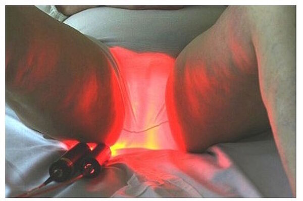
The light emitting fabrics technology can be associated with different light sources and can deliver different illumination modalities, in compliance with regulatory constraints (such as the Levulan® in the US, which requires illumination at 417 nm) and clinical constraints.
1 Vignion-Dewalle, A.S., et al., A software for analyzing and comparing the light sources available for PDT in dermatology. EuroPDT Munich, 2017.
2 Vignion-Dewalle, A.S., et al., Comparison of three light doses in the photodynamic treatment of actinic keratosis using mathematical modeling. J Biomed Opt, 2015. 20(5): p. 58001.
3 Mordon, S., et al., Light emitting fabric technologies for photodynamic therapy. Photodiagnosis Photodyn Ther, 2015. 12(1): p. 1-8
4 Phase II study evaluating the non-inferiority of the Flexitheralight device compared to conventional phototherapy (PDT): n° 2013-A01096-39 – ID clinical trial: NCT03076918
5 (www.phosistos.com)
6 Phase II study evaluating the non-inferiority of the Phosistos device compared to conventional phototherapy (PDT): n° 2016-A00010-51 – ID clinical trial: NCT03076892
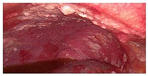
PRODYNOV project is a multidisciplinary approach that aims to enable intraperitoneal photodynamic therapy (PDT) of peritoneal metastases of epithelial ovarian cancer (EOC).
Background
EOC management remains association of complete cytoreductive surgery in combination with platinum-based chemotherapy and targeted therapies if indicated. Despite this management, 60% of patients in remission will develop peritoneal recurrences. This high peritoneal recurrence rate raises the issue of microscopic peritoneal metastases management and requires the development of additional locoregional treatment strategies.
An option to improve the completion of cytoreductive surgery may be the use of PDT. Photodynamic therapy’s ability to treat superficial lesions disseminated on large area makes it an excellent candidate to insure destruction of microscopic peritoneal metastases.
This treatment may be associated with fluorescence guided surgery using the same photosensitizer to improve the completion of cytoreductive surgery.
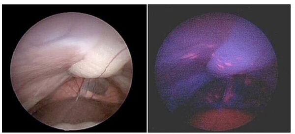
Photodynamic Therapy
PDT is an effective technique that has already been applied in other medical indications. After administration of a photosensitizer (PS) that accumulates in cancer cells, illumination with a light of adequate wavelength induces a photochemical reaction to tissue oxygen leading to reactive oxygen species (ROS) production and a cytotoxic phenomenon. PDT ability to treat superficial lesions over a large area makes it an excellent candidate for destruction of microscopic residual disease.
Theoretically PDT has the advantage of a double selectivity: photosensitizer selectivity for the tumor and selectivity of the wavelength that activates the photosensitizer, but the development of intraperitoneal PDT has been limited because of poor tolerance related to the lack of specificity of photosensitizers and the proximity of intraperitoneal organs. Recent approaches targeting folate receptors which are highly specific for EOC could resolve this limitation. The French team Réactions et Génie des Procédés (UMR 7274 CNRS – University of Lorraine, Nancy, France) is expert in the field of specific photosensitizer design and synthesis.
PDT is also interesting because it’s based on different biological mechanisms and may offer a cytotoxic response that involves different pathways than chemotherapy, in case of chemoresistance. Furthermore, in addition to direct cytotoxicity, PDT can stimulate a delayed immune response and be responsible for immunotherapy (abscopal effect). Recent studies demonstrate that PTD may change the secretome of tumor cells in favor of a secretome that activates the mitochondrial metabolism of immune cells. The team CNRS UMR 8161 (Institut de Biologie de Lille, France) is working on this topic.
PDT could offer well-tolerated and effective adjuvant therapy of the peritoneum after surgical removal of visible tumors.
Where do we stand?
Preclinical studies have been performed in vitro on murine and human cell lines of EOC and in vivo with a preclinical model of peritoneal carcinomatosis.
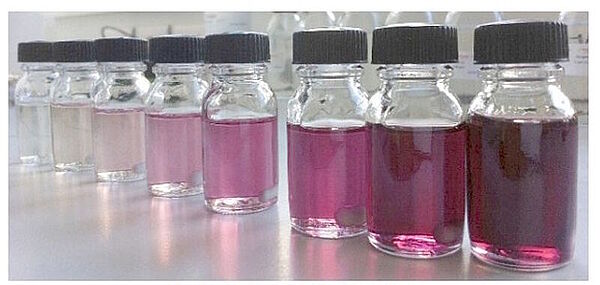
Tissue quantification of the PS showed specific incorporation of the folate-targeted PS within tumor tissue. Specificity for ovarian cancer metastases is better than previously reported with others photosensitizers (Tumor-to-normal tissue ratio 9.6). We could detect specific fluorescence in vitro and in vivo on peritoneal metastases. Folic-acid targeted PDT allows to obtain human EOC cells death in vitro.
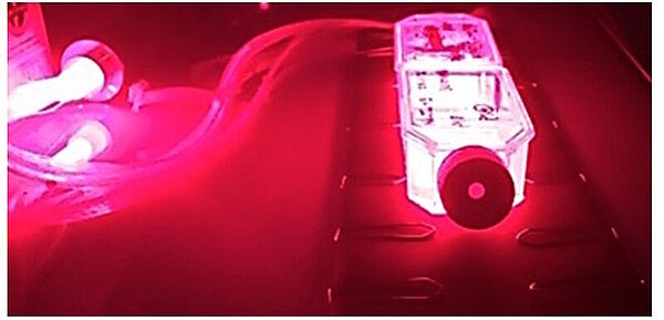
Another aspect of photodynamic therapy development is the illumination using an optimal scheme and a light administration monitoring. Innovative illumination solutions are available, as textile light diffusers which offer the possibility to apply a homogenous distribution of light on large surface area, as parietal peritoneum or direct and cylindrical diffusing fibres which are easy to handle and allow reaching spaces that are difficult to attain.
Context and Challenges
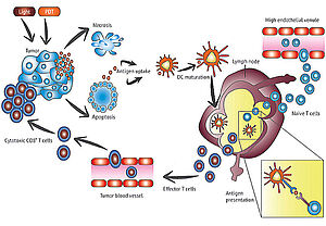
The use of PDT in the field of oncology has already proved its worth and has been the subject of growing interest in recent years. Today, there is a real rational for thinking that PDT could impact the immune response, in favour of immunoactivation. This property would be a considerable asset in the management of cancers if we take into account the success of certain anti-tumour immunotherapies (anti-PD1, PDL1, CTLA-4, etc.), particularly in the last 3 years.
Within the ONCOTHAI team, the ImmunoPDT team led by Pr Nadira DELHEM works in close association with the PhysicoPDT team led by Dr Anne-Sophie Dewalle, and in collaboration with clinicians from the Lille CHRU and the Centre Oscar Lambret. The five major projects developed within the team aim to better understand the impact of PDT on the immune response by evaluating more specifically the mechanisms involved in the immuno-modulation of the immune system by PDT.
The realization of these projects requires a double expertise, that acquired historically by the team "Immunoregulation of Viro-Induced Cancers" led by Pr Nadira Delhem, in the fields of immunosurveillance and cancers' immunotherapy, and that acquired by "Physico PDT" team in the field of lasers and photodynamic therapy. The synergy of these skills will enable us to understand the role of PDT in immune regulation upstream, in order to propose innovative protocols for the treatment of cancers.
Objectives
The ONCOTHAI team, in collaboration with the clinical departments of Gynecology, Digestive Transplantation, Neurosurgery, Dermatology and Pneumology of the Lille CHRU, has started five ambitious projects which aimed to evaluate, in parallel, the efficacy of PDT as an anti-tumor treatment and the impact of PDT on the regulation of the immune response. This translational work is taking place in two areas: clinical and research. In terms of research, the immunoPDT team proposes to develop fundamental projects further upstream of translational research, aiming to understand at the biological and molecular level, the impact of PDT on both tumour cells and immune cells [IM-PRODYNOV (ovarian carcinosis) and HEPATOCAR (hepatocarcinoma) projects].
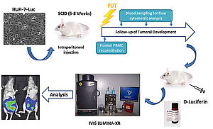
In addition, the ImmunoPDT team has developed high-performance methodological tools and techniques for the in vivo evaluation of new anti-tumor therapeutic strategies.
In particular, the team has developed innovative "custom" humanized mouse models that combine xeno-transplantation of human tumors expressing luciferase with reconstitution with a human immune system.
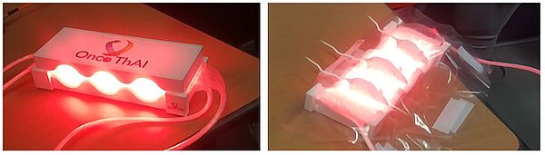
These models, which are easily exploitable in small animal imaging, make it possible to develop anti-tumor strategies in a context very close to human physiopathology.
In order to evaluate in these in vivo models the efficacy of PDT and its impact on a human immune system, unit U1189 has developed innovative devices for in vivo illumination of mice.
At the clinical level, the ImmunoPDT team is developing more translational research projects aimed at characterizing the effects of PDT, combined with surgery or immunotherapy, on the phenotype and activation of immune cells (immunomonitoring) and on the regulation of the immune response (regulatory T lymphocytes, tolerogenic DC, tumor exosomes, etc.). All of these studies are carried out using samples from patients included in clinical trials or ancillary studies [IM-INDYGO (glioblastoma), IMCUTALA (melanoma) and IMPALA (mesothelioma) projects].
Where are we with our research?
Thanks to the financial support of SATT Nord de France and SATT Grand-Est, a patent on a new photosensitizer (PS), specifically targeting peritoneal ovarian carcinosis, has been filed. The photosensitizer (PS-FOL) presented here is the product of an original synthesis carried out by the team of the Reaction and Process Engineering Laboratory (LRGP - UMR 7274 CNRS - University of Lorraine). The objective of this synthesis is to associate the PS with an addressing molecule (folic acid) targeting FR with a strong affinity. We cannot further detail the nature of this compound for confidentiality reasons.
Our main objective was to evaluate PDT in this indication and its impact on the immune response. Initial in vitro results showed an excellent efficacy of PDT on human ovarian carcinoma cell lines.
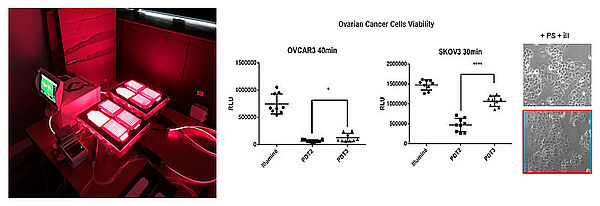
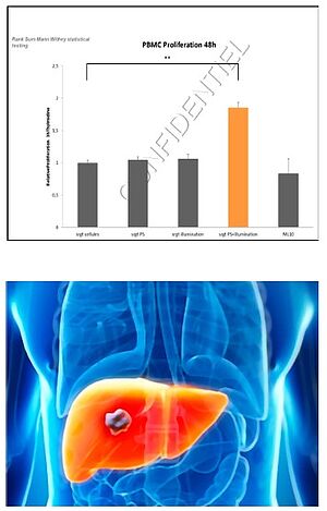
Furthermore, PDT also has a beneficial effect on the immune system. The observed results show that tumour cells subjected to PDT produce immunostimulatory factors by inducing an increase in the proliferation of human immune cells.
PDT also induces a decrease in the production of pro-inflammatory cytokines by tumour cells and an increase in cytokines that are favourable to the survival, proliferation and activation of immune cells, mainly for CD4+ T and CD8+ T lymphocytes, such as the cytokines IL2, IFN-gamma.
In the context of hepatocellular carcinoma (HCC), we were able to obtain very promising initial results thanks to international thesis funding. The main objective of this study is to understand, in the context of HCC, the direct and indirect effects of 5-Aminolevulinic Acid (5-ALA)-mediated HCC.
In this context, hepatic cancer cell lines have been used in a first step: HuH7 (overexpressing p53), Hep3B (partial deletion of p53) and HepG2 (expressing wild p53) and in a second step primary HCC cells from surgical excision will be used.
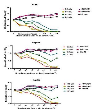
Our preliminary results showed that PDT can be effective against the three different HCC cell lines because they express the key enzymes responsible for the conversion of the pro-drug 5-ALA into Porphyrin IX (PpIX), the final PS.
Thus, PDT treatment with 5-ALA in these cell lines induces cell death that is dependent on the dose of 5-ALA and the duration of illumination.
Interestingly, we also observed that these three cell lines respond variably to 5-ALA PDT in a pattern that appears to be related to the p53 expression profile.
Moreover, thanks to a very close collaboration network with clinicians from the CHRU of Lille and the Centre Oscar Lambret and thanks to the logistic and financial support of industrials (Novartis, BMS and MSD), we have initiated two new projects in the framework of Melanoma and Mesothelium.
Immunotherapies, such as anti-PD-1 antibodies, have emerged in recent years as a major therapeutic tool in many cancers. Thus, the advantages provided by both immunotherapy and PDT have led us to propose a combination of the two treatments in order to improve the local anti-tumor effect in patients with melanoma with skin metastases, or with mesothelium.
Based on a rational synergistic effect of PDT and immune checkpoint inhibitors, we hope to induce a strong anti-tumor immune response, in the absence of cumulative toxicities, leading to a better outcome for these patients.